![]() Figure 1 of
Al-Ghoul, Mol Vis 1999;
5:6.
Figure 1 of
Al-Ghoul, Mol Vis 1999;
5:6.
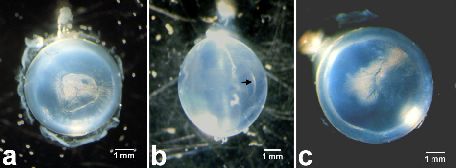
Figure 1. Light micrographs of whole, fixed RCS rat lenses.
(A) The posterior aspect of a 3 month old lens showing a PSC plaque. Because internalization of the PSC commences between 8 to 10 weeks, the plaque is only covered by about 0.2 mm of new growth and consequently still appears very prominent. (B) An equatorial view of a 6 month old lens demonstrating the internalized plaque (arrow) covered by approximately 0.5 mm of new growth. (C) Posterior view of a six month old lens showing an abnormal suture pattern. Instead of the typical inverted Y pattern, this lens had a smaller, 5-branched suture pattern.