![]() Figure 8 of
Thulin, Mol Vis 1999;
5:40.
Figure 8 of
Thulin, Mol Vis 1999;
5:40.
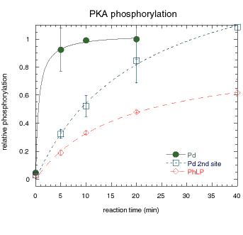
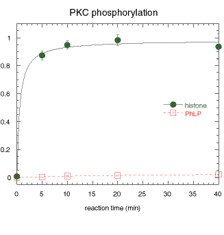
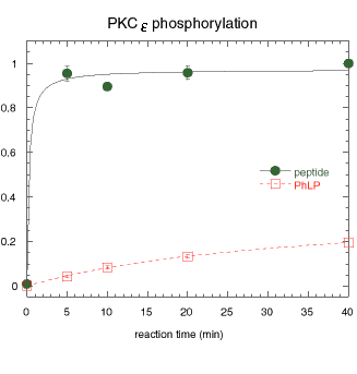
Figure 8. Phosphorylation of PhLP by PKA and PKC isoforms
Phosphorylation of PhLP, Pd, histone and peptide substrates by PKA and PKC isoforms. Phosphorylation conditions are specified in Methods. Error bars represent standard deviation of duplicate or triplicate samples.
A. Protein kinase A catalytic subunit was used to phosphorylate purified recombinant Pd or PhLP in vitro and the 32P incorporated determined by phosphorimager analysis of SDS gels. The relative amount of phosphorylation is shown for the high-efficiency site that has been shown to be Ser73 of Pd (filled circles), for a second less efficient site on Pd which results in the appearance of a second band (open squares), and for PhLP (open diamonds).

B. Protein kinase C from rat brain (a mixture of isoforms [alpha], ß, and [gamma]) was used to phosphorylate a standard substrate (histone, filled circles) and recombinant PhLP (open squares).

C. Recombinant protein kinase C[epsilon] was used to phosphorylate a peptide substrate (filled circles) and recombinant PhLP (open squares).
