![]() Figure 7 of
Thulin, Mol Vis 1999;
5:40.
Figure 7 of
Thulin, Mol Vis 1999;
5:40.
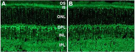
Figure 7. Specificity of PhLP immunolocalization
A. Fluorescein isothiocyanate staining shows localization of PhLP using the anti-PhLP N-terminus/GST fusion antibody. B. Staining in the same section is somewhat reduced overall but relative intensity of staining in the photoreceptor inner segments (top of panels) is maintained using antibody that has been depleted with 25 µg/ml recombinant Pd.