![]() Figure 6 of
Thulin, Mol Vis 1999;
5:40.
Figure 6 of
Thulin, Mol Vis 1999;
5:40.
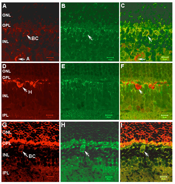
Figure 6. Immunolocalization of PhLP compared with that of calretinin, calbindin and recoverin
A. Rhodamine staining indicates localization of calretinin to amacrine cells (arrow) and bipolar cells (arrow). B. FITC staining indicates localization of PhLP in the same section (arrow). C. Double staining (yellow) indicates colocalization of calretinin and PhLP in bipolar cells but not amacrine cells (arrows). D. Rhodamine staining shows localization of calbindin, reported to localize to horizontal cells (arrow). E. FITC staining shows localization of PhLP in the same section. F. Double staining indicates the lack of significant colocalization of calbindin and PhLP in horizontal cells (arrow). G. Rhodamine staining localizes recoverin to photoreceptor cells and bipolar cells (arrow). H. FITC staining localizes PhLP in the same section (arrow). I. Double staining shows colocalization of recoverin and PhLP in bipolar cells (arrow), but not in photoreceptor cells.