![]() Figure 5 of
Thulin, Mol Vis 1999;
5:40.
Figure 5 of
Thulin, Mol Vis 1999;
5:40.
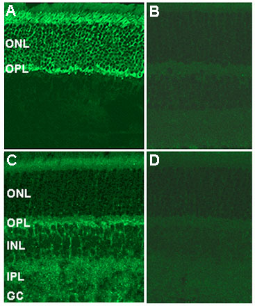
Figure 5. Immunocytochemical localization of Pd and PhLP in mouse retinal sections
A. Fluorescein isothiocyanate (FITC) staining indicates the localization of Pd to the photoreceptor cells, including the inner segments (top), outer nuclear layer (ONL), and outermost parts of the outer plexiform layer (OPL). C. FITC staining shows localization of PhLP to the region near the OPL, the inner nuclear layer (INL), inner plexiform layer (IPL), and photoreceptor inner segments (top). Specificity of staining was demonstrated by competing away signal with preincubation of the primary antibody with 25 µg/ml recombinant Pd (B) or PhLP N-terminus/GST fusion protein (D).