![]() Figure 2 of
Thulin, Mol Vis 1999;
5:40.
Figure 2 of
Thulin, Mol Vis 1999;
5:40.
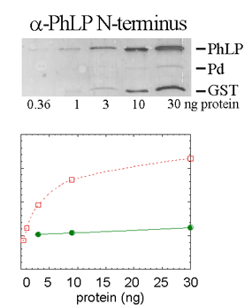
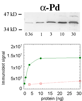
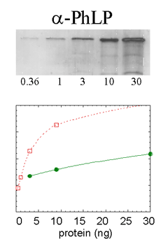
Figure 2. Specificity of the affinity-purified antibodies raised against phosducin and phosducin-like protein
An equal mixture of recombinant PhLP, Pd, and GST were electrophoresed using the amounts of protein indicated at the bottom of A-C (0.36-30 ng of each protein). Immunoblots were probed with anti-phosducin (A), anti-PhLP (B), and anti-PhLP N-terminus/GST fusion protein (C). See Methods for antibody details. Shown with the blots are graphs of immunoblot signal for Pd (filled circles, solid line interpolated) and PhLP (open squares, dotted line). Specificity was quantified by comparing the slopes of signals in the linear range (first three points).
A. Anti-phosducin.
B. Anti-PhLP.
C. Anti-PhLP N-terminus/GST fusion protein.