![]() Figure 1 of
Thulin, Mol Vis 1999;
5:40.
Figure 1 of
Thulin, Mol Vis 1999;
5:40.
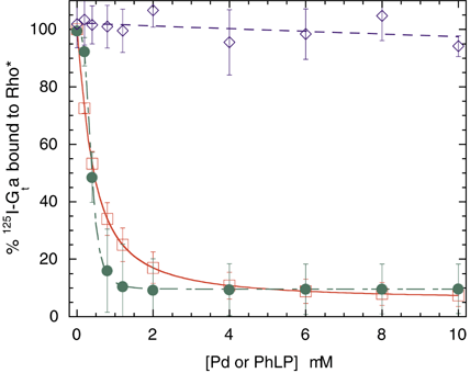
Figure 1. Phosducin (Pd) and PhLP inhibit Gt binding to light-activated rhodopsin (Rho*)
Light-induced binding of 125I-Gt[alpha] (0.2 µM) and Gtß[gamma] (0.2 µM) to urea-stripped rod outer segment membranes (1.0 µM Rho) was carried out in the presence of Pd (green, filled circles) or PhLP (red, open squares) at the concentrations indicated (see Methods). Ovalbumin shown as negative control (blue, open diamonds). Error bars represent the first standard deviation of data from three separate experiments. 100% is 0.07-0.1 pmol of 125I-Gt[alpha]bound/pmol of Rho*. Lines represent non-linear least squares fit of the data to the equation: Fraction bound = (1 - X)/(1 + [Pd]/K1/2) + X, where X is the fraction remaining bound at saturating the concentration of Pd and K1/2 is the concentration of Pd at which half maximal inhibition occurred. K1/2 values were 0.37 µM for Pd and 0.41 µM for PhLP.