![]() Figure 4 of
Seigel, Mol Vis 1999;
5:4.
Figure 4 of
Seigel, Mol Vis 1999;
5:4.
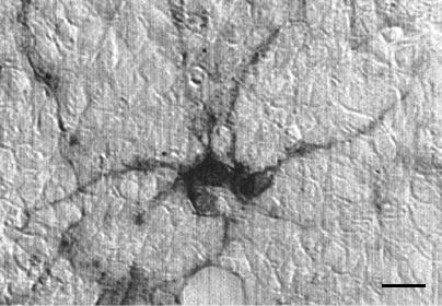
Figure 4. R28 retinal cells immunoreactive to Thy 1.1
The photograph depicts R28 cells immunoreactive for Thy 1.1, a retinal ganglion cell marker. Scale bar is 5 µm.
R28 cells were grown in DMEM with 10% calf serum, 1 mM L-glutamine, and 50 µg/ml gentamicin. For Thy 1.1 staining, cells were fixed for 10 min at room temperature in 2% paraformaldehyde and permeabilized in 0.25% Triton X-100 for 5 min. After a rinse in phosphate-buffered saline (PBS), cells were incubated for 1 h with 10 µg/ml anti-Thy 1.1 antibody (Chemicon, Temecula, CA). After rinsing 3 times for 5 min in PBS, cells were incubated with biotinylated anti-mouse immunoglobulin (Vector Laboratories, Burlingame, CA) for 60 min. Cells were equilibrated in Tris-buffered saline (50 mM Tris-HCl, pH 7.6, 0.9% NaCl) and incubated for 20 min with horseradish peroxidase-conjugated avidin (Elite kit, Vector Laboratories). The cells were rinsed in 0.05 M Tris and developed with a diaminobenzidine kit (Pierce, Rockford, IL) and the brown/black reaction product visualized by light microscopy. Negative controls consisted of incubations in control serum without primary antibody, and did not generate reaction product.