![]() Figure 2 of
Seigel, Mol Vis 1999;
5:4.
Figure 2 of
Seigel, Mol Vis 1999;
5:4.
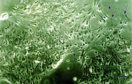
Figure 2. Dissociated E17 rat retina in vitro
Embryonic day 17 rats were enucleated and the neural retina dissected free of pigment epithelium. A single cell suspension was generated by treating the tissue for 10 min at 37 °C with 0.6% trypsin in 1 mM EDTA. The tissue was rinsed with phosphate-buffered saline and plated into 35 mm dishes with Dulbecco's Modified Eagle's Medium containing 1 mM L-glutamine, 10% fetal calf serum and 50 µg/ml gentamicin. Dishes were placed in a humidified chamber with 5% CO2, 95% air at 37 °C.
The E17 culture was photographed after 5 days in vitro. Note the neuritic processes (arrowheads), as well as flat cells (arrows). Scale bar is 10 µm.