![]() Figure 1 of
Seigel, Mol Vis 1999;
5:4.
Figure 1 of
Seigel, Mol Vis 1999;
5:4.
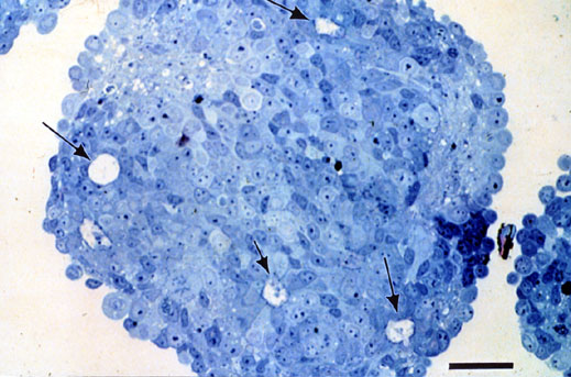
Figure 1. Chick Retinal Reaggregate Culture
Embryonated chicken eggs of C/E genotype from Spafas (Norwich, CN) were allowed to develop in an egg incubator for 7 days. The chick embryo was removed and the neural retinae were dissected free of pigmented epithelium and optic nerve. A single cell suspension was generated by treating the tissue for 10 min at 37 °C with 0.6% trypsin in 1 mM EDTA. The tissue was rinsed with phosphate-buffered saline and seeded into 35 mm suspension-culture dishes at 5 x 10⁶ cells per dish with Dulbecco's Modified Eagle's Medium with 1 mM L-glutamine, 10% fetal calf serum and 50 µg/ml gentamicin. Dishes were placed on a Junior Orbit Shaker set at 70 rpm in a humidified chamber with 5% CO2, 95% air at 37 °C.
The photo depicts a retinal reaggregate after 7 days in vitro. Rosettes organized around central lumen are visible (arrows). Scale bar is 10 µm.