![]() Figure 4 of
Stewart-DeHaan, Mol Vis 1999;
5:37.
Figure 4 of
Stewart-DeHaan, Mol Vis 1999;
5:37.
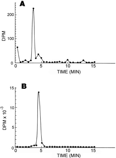
Figure 4. HPLC separation of 3H-glutathione from lens homogenate compared to the standard 3H-glutathione injected into the rat
Lenses were removed after sacrifice of the rat at 30 min after injection of 3H-glutathione, weighed and homogenized in 1.0 ml PEDTA. 0.1 ml of 69% HClO4 was then added to deproteinize the homogenate. An aliquot of the 3H-glutathione injected was also taken for HPLC using a C18 Bondapak column as described in the text. The volumes used for injection were 20 µl of deproteinized lens homogenate (total volume 1.09 ml) and 5 µl glutathione (diluted with 0.1 volumes of 60% HClO4). The total radioactivity in the aliquots used was 6432 dpm/ 100 µl homogenate injected, and 1.029 x 10⁷ dpm for 200 µl of standard glutathione which was injected. Fractions (0.5 ml) were collected at a flow rate of 1 ml/min into scintillation vials and counted to establish the elution profile. As can be seen, the radioactive glutathione appears to elute in fraction 9.