![]() Figure 2 of
Martin, Mol Vis 1999;
5:36.
Figure 2 of
Martin, Mol Vis 1999;
5:36.
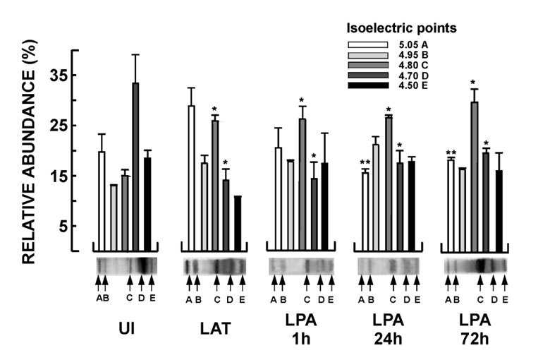
Figure 2. Relative abundance of rabbit corneal GAP-43 isoforms focusing at pI 5.05, 4.95, 4.80, 4.70, and 4.50
The pI 4.7 GAP-43 isoform (band D) was most abundant in uninfected rabbits (UI). Comparing UI rabbits with the other groups, band C (pI 4.80) was more abundant and band D (pI 4.70) was decreased in all infected conditions (*). After lysophosphatidic acid (LPA) iontophoresis, the collective posttranslational modification of GAP-43 in latently-infected rabbits was increased because the relative abundance of the most basic GAP-43 isoform (band A, pI 5.05) was decreased at 24 h and 72 h (**). Beneath each group, bands (A-E) are shown in a gel exemplary of those used for densitometric analysis. Values are densitometric averages ± standard error of four separate determinations (corneas). One-way ANOVA with post-hoc Newman-Keuls tests show significant differences between treatment groups (p<0.05). The (*) indicates significant differences between uninfected corneas (UI) and all other experimental groups. The (**) indicates significant differences between the latently-infected corneas (LAT) and the LPA-treated, latently-infected corneas.