![]() Figure 1 of
Martin, Mol Vis 1999;
5:36.
Figure 1 of
Martin, Mol Vis 1999;
5:36.
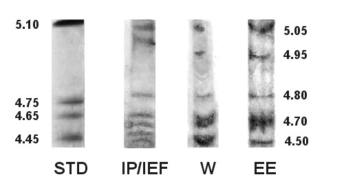
Figure 1. Three methods of GAP-43 identification
In the first method, proteins were immunoprecipitated (IP) with monoclonal antibody 91E12 and applied to isoelectric focusing (IEF) gels (IP/IEF). In the second method, proteins were applied to IEF gels and transferred to nitrocellulose membranes for Western blotting with monoclonal antibody 91E12 and chemiluminescent detection (W). In the third method, proteins were electroeluted from the 43 kDa region of sodium dodecyl sulfate polyacrylamide gels, applied to IEF gels, and silver stained (EE). The proteins of interest had isoelectric points of 5.05, 4.95, 4.80, 4.70, and 4.50. In some preparations, two bands focused near pI 4.70 and 4.50. The isoelectric points of standard proteins (STD) are noted at the left and those of GAP-43 isoforms are noted at the right.