![]() Figure 9 of
Winkler, Mol Vis 1999;
5:32.
Figure 9 of
Winkler, Mol Vis 1999;
5:32.
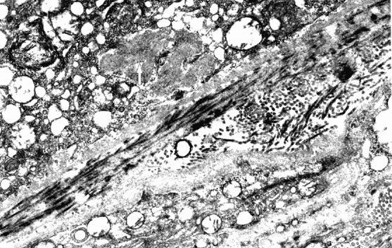
Figure 9. Sub-retinal pigment epithelial deposits in a mouse model of protoporphyria
Protoporphyric mouse model exposed to blue light demonstrates sub-retinal pigment epithelium fibrillogranular deposit (white arrow) with fibrils measuring up to 16 nm with a periodicity of 13 nm.