![]() Figure 7 of
Winkler, Mol Vis 1999;
5:32.
Figure 7 of
Winkler, Mol Vis 1999;
5:32.
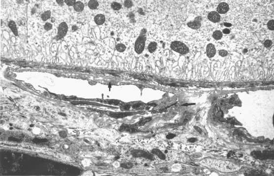
Figure 7. Choriocapillaris in a mouse model of protoporphyria
In the mouse model of protoporphyria with approximately a 10-fold increase in protoporphyrin IX and exposure to blue light (380-430 nm, 14 µW/cm2), a time and light dependent increase in choriocapillary and subretinal pigment epithelial basal laminar-like deposits are demonstrated (see arrows).