![]() Figure 4 of
Hageman, Mol Vis 1999;
5:28.
Figure 4 of
Hageman, Mol Vis 1999;
5:28.
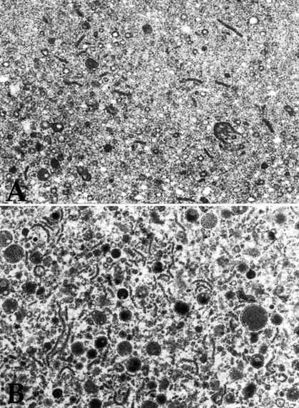
Figure 4. High magnification electron micrographs of the second drusen phenotype
High magnification transmission electron micrographs depicting the accumulation of curvilinear profiles and spherical elements in some drusen of the second phenotype (Figure 3). Note the increase in heterogeneity between A and B. In some cases, drusen of this phenotype possess spherical elements, of which some are membrane-bounded and all are electron dense (A and B).