![]() Figure 8 of
Green, Mol Vis 1999;
5:27.
Figure 8 of
Green, Mol Vis 1999;
5:27.
Figure 8. Choroidal neovascularization
A. Area where a choroidal artery (asterisk) extends through a defect in Bruch's membrane and into a plane between BlamD (arrowhead) and the remainder of Bruch's membrane. (periodic acid-Schiff, x16)
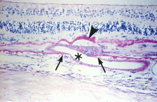
Figure 8A reprinted with permission, from: Green WR, Enger C. Age-related macular degeneration histopathologic studies. The 1992 Lorenz E. Zimmerman Lecture. Ophthalmology 1993; 100:1519-35.
B. Area where a choroidal artery (A) and vein (V) extends through a defect in Bruch's membrane (between arrows) and into a plane between BlamD with RPE and the remainder of Bruch's membrane. (periodic acid-Schiff, x160)
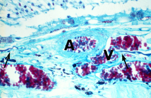
Figure 8B reprinted with permission, from: Green WR, Enger C. Age-related macular degeneration histopathologic studies. The 1992 Lorenz E. Zimmerman Lecture. Ophthalmology 1993; 100:1519-35.