![]() Figure 7 of
Green, Mol Vis 1999;
5:27.
Figure 7 of
Green, Mol Vis 1999;
5:27.
Figure 7. Early choroidal neovascularization
Examples of choroidal neovascularization with capillaries (asterisks) extending through defects in Bruch's membrane (between arrows) and into the plane between RPE and Bruch's membrane (periodic acid-Schiff, x40).
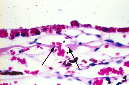
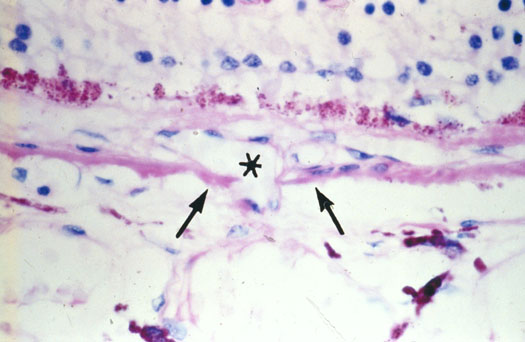
Figure 7 (lower) reprinted with permission, from: Green WR, Enger C. Age-related macular degeneration histopathologic studies. The 1992 Lorenz E. Zimmerman Lecture. Ophthalmology 1993; 100:1519-35.