![]() Figure 6 of
Green, Mol Vis 1999;
5:27.
Figure 6 of
Green, Mol Vis 1999;
5:27.
Figure 6. Early choroidal neovascularization
Examples of thin subretinal pigment epithelial fibrovascular membranes (asterisks) located between the layer of basal laminar deposit (arrows) and Bruch's membrane. The retinal pigment epithelium is intact, with moderate photoreceptor cell degeneration present (Van de Grift, x40)
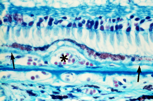
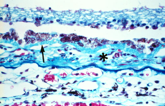
Figure 6 reprinted with permission, from: Green WR, Enger C. Age-related macular degeneration histopathologic studies. The 1992 Lorenz E. Zimmerman Lecture. Ophthalmology 1993; 100:1519-35.