![]() Figure 3 of
Green, Mol Vis 1999;
5:27.
Figure 3 of
Green, Mol Vis 1999;
5:27.
Figure 3. Basal linear deposit
A. Ultrastructural appearance of a 6.4 µm-thick basal linear deposit composed of vesicular material (asterisks) located between the retinal pigment epithelial basal lamina (arrowhead) and the remainder of Bruch's membrane. A 2.2 µm-thick layer of wide-spaced collagen (basal laminar deposit, between arrows) is present internal to the retinal pigment epithelial basal lamina (arrowhead). x15,000.
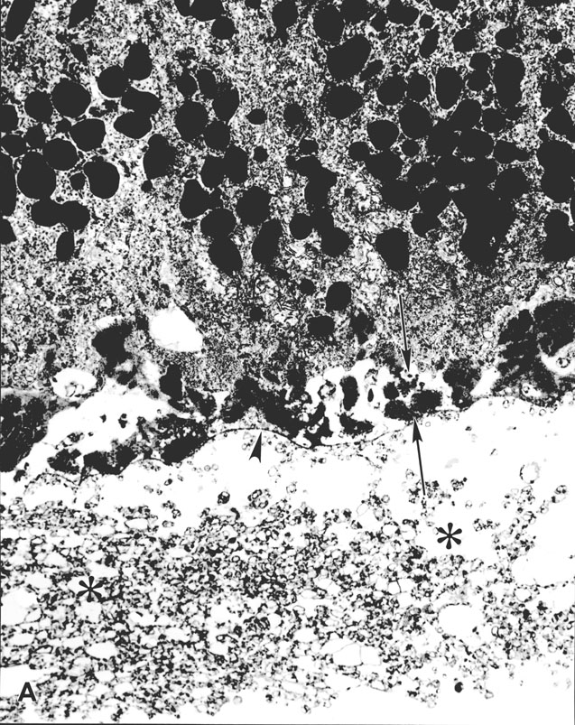
Figure 3A reprinted with permission, from: Green WR, Enger C. Age-related macular degeneration histopathologic studies. The 1992 Lorenz E. Zimmerman Lecture. Ophthalmology 1993; 100:1519-35.
B. Higher power view of vesicular material in basal linear deposit (x40,000).
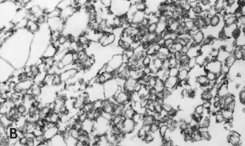
Figure 3B reprinted with permission, from: Green WR, Enger C. Age-related macular degeneration histopathologic studies. The 1992 Lorenz E. Zimmerman Lecture. Ophthalmology 1993; 100:1519-35.
C. Higher power view of basal laminar deposit with foci of wide-spaced collagen located internal to the RPE basal lamina (arrowhead). (x40,000)
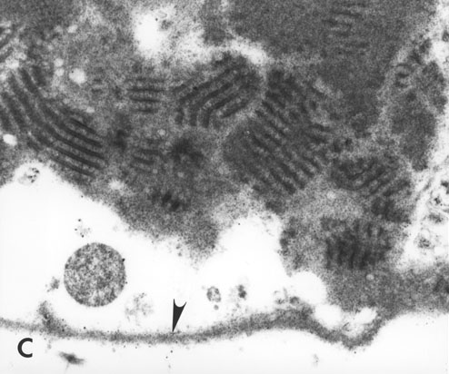
Figure 3C reprinted with permission, from: Green WR, Enger C. Age-related macular degeneration histopathologic studies. The 1992 Lorenz E. Zimmerman Lecture. Ophthalmology 1993; 100:1519-35.