![]() Figure 2 of
Green, Mol Vis 1999;
5:27.
Figure 2 of
Green, Mol Vis 1999;
5:27.
Figure 2. Basal laminar deposit
A. Phase contrast appearance of BlamD with serrated inner margin. (x9,900).

B. Thick layer of basal laminar deposit is located between the plasma (PM) and basal lamina (arrowhead) of the RPE and consists of membrano-granular material (circle) and foci of wide-spaced collagen (arrows). Splitting (asterisk) has occurred in this new, thick layer. Brackets mark the elastic tissue layer of Bruch's membrane. (x50,000).
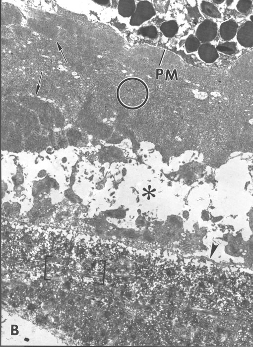
Figure 2B reprinted with permission, from: Green WR, Key SN 3d. Senile macular degeneration: a histopathologic study. Trans Am Ophthalmol Soc 1977; 75:180-254.
C. Membrano-granular material (x29,000). D. Wide-spaced collagen (x29,000).
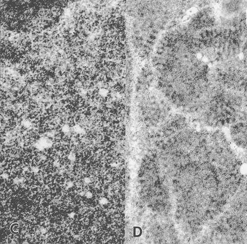
Figures 2C-D reprinted with permission, from: Green WR, Key SN 3d. Senile macular degeneration: a histopathologic study. Trans Am Ophthalmol Soc 1977; 75:180-254.