![]() Figure 19 of
Green, Mol Vis 1999;
5:27.
Figure 19 of
Green, Mol Vis 1999;
5:27.
Figure 19. Retinal vascularization of disciform scar
Low (A) and higher power view (B) of a two-component, 289 µm maximal thickness, disciform scar with marked to total atrophy of the photoreceptor cell layer. The subretinal component (single asterisk) is vascularized by a vessel (arrow) from the retina. A 7.1 µm-thick layer of basal laminar deposit (arrowhead) separates the subretinal (single asterisk) and intra-Bruch's membrane (double asterisk) components of the scar. The intra-Bruch's membrane component is vascularized by an artery and vein from the choroid at a different level. Periodic acid-Schiff: A, x45; B, x155.
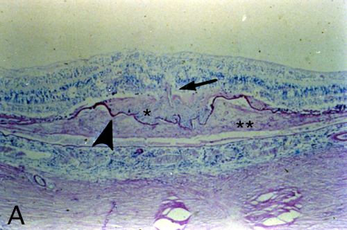
Figure 19A reprinted with permission, from: Green WR, Enger C. Age-related macular degeneration histopathologic studies. The 1992 Lorenz E. Zimmerman Lecture. Ophthalmology 1993; 100:1519-35.
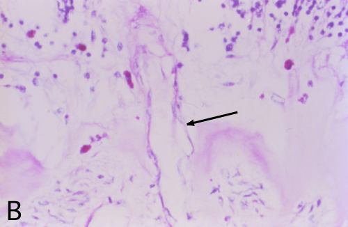
Figure 19B reprinted with permission, from: Green WR, Enger C. Age-related macular degeneration histopathologic studies. The 1992 Lorenz E. Zimmerman Lecture. Ophthalmology 1993; 100:1519-35.