![]() Figure 18 of
Green, Mol Vis 1999;
5:27.
Figure 18 of
Green, Mol Vis 1999;
5:27.
Figure 18. Choroidal neovascularization
A. A two-component, 100 µm maximal thickness, disciform scar with a small subretinal component (single asterisk) and a small area of serous detachment of the macula (arrowhead). There is moderate to total loss of the photoreceptor cell layer over the scar, which is most marked over the thicker intra-Bruch's membrane component (double asterisk). (periodic acid-Schiff; x35).
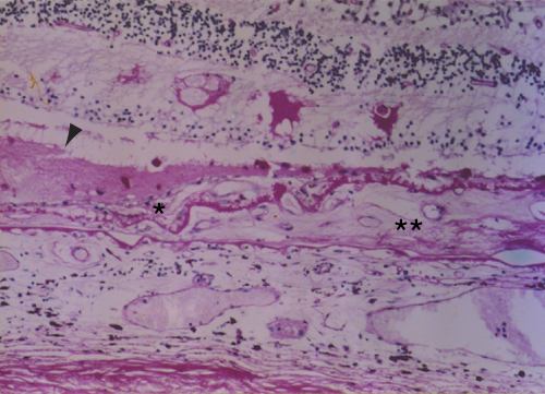
Figure 18A reprinted with permission, from: Green WR, Enger C. Age-related macular degeneration histopathologic studies. The 1992 Lorenz E. Zimmerman Lecture. Ophthalmology 1993; 100:1519-35.
B. Higher power view of the vascularized intra-Bruch's membrane (BM) component (double asterisk) where the basal laminar deposit has a typical brush-like inner surface and a defect (between arrows) where a capillary (arrowheads) extends from the intra-Bruch's membrane component to the thin subretinal component (Van de Grift; x360).
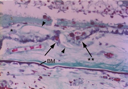
Figure 18B reprinted with permission, from: Green WR, Enger C. Age-related macular degeneration histopathologic studies. The 1992 Lorenz E. Zimmerman Lecture. Ophthalmology 1993; 100:1519-35.