![]() Figure 17 of
Green, Mol Vis 1999;
5:27.
Figure 17 of
Green, Mol Vis 1999;
5:27.
Figure 17. Disciform scar with tear of RPE and BlamD
A. A tear of RPE and BlamD (between arrows) is present at the nasal margin of a two-component disciform scar. Periodic acid-Schiff, x2.5.
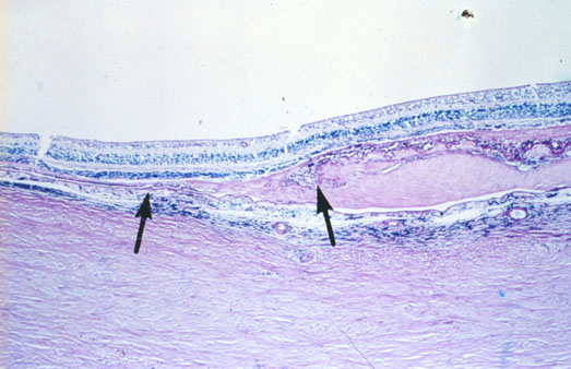
Figure 17A reprinted with permission, from: Green WR, Enger C. Age-related macular degeneration histopathologic studies. The 1992 Lorenz E. Zimmerman Lecture. Ophthalmology 1993; 100:1519-35.
B. Higher power view of the RPE/BLamD tear where the nasal margin of the tear has pulled under temporally (arrows) and the area of the tear is filled in by fibrous tissue. Periodic acid-Schiff, x25.
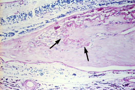
Figure 17B reprinted with permission, from: Green WR, Enger C. Age-related macular degeneration histopathologic studies. The 1992 Lorenz E. Zimmerman Lecture. Ophthalmology 1993; 100:1519-35.