![]() Figure 14 of
Green, Mol Vis 1999;
5:27.
Figure 14 of
Green, Mol Vis 1999;
5:27.
Figure 14. Sub-RPE disciform scar
Example of thin, non-vascularized, subretinal pigment epithelial fibrocellular disciform scars (asterisk) located between basal laminar deposit (arrow) and the remainder of Bruch's membrane. The retinal pigment epithelium and photoreceptor cell layer are atrophic over the scar. Periodic acid-Schiff, x25.
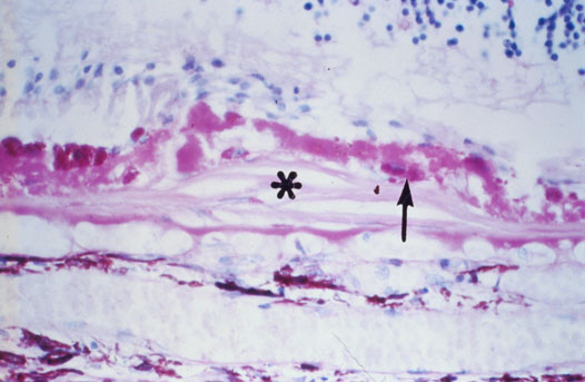
Figure 14 reprinted with permission, from: Green WR, Enger C. Age-related macular degeneration histopathologic studies. The 1992 Lorenz E. Zimmerman Lecture. Ophthalmology 1993; 100:1519-35.