![]() Figure 13 of
Green, Mol Vis 1999;
5:27.
Figure 13 of
Green, Mol Vis 1999;
5:27.
Figure 13. Indocyanine green hot spot
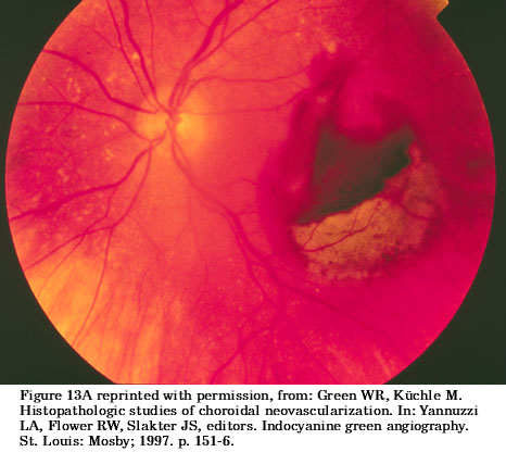
A. Preoperative clinical photograph demonstrating subretinal hemorrhage in the macular area. The inferior portion of the extravasated blood has lost its hemoglobin, resulting in the color change.
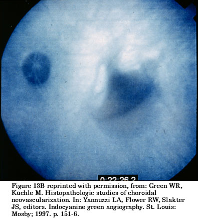
B. Late-phase indocyanine green angiogram demonstrating blocked fluorescence from the thicker layers of hemorrhage. A more extensive superior area of hyperfluorescence corresponds to an area of occult CNV. In addition, a linear area of more intense hyperfluorescence just nasal to the blocked fluorescence is consistent with a site of neovascularization ("hot spot").
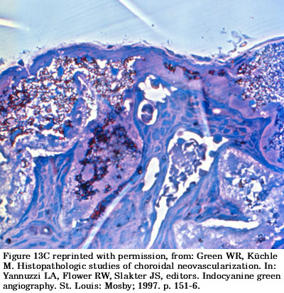
C. Submacular membranectomy specimen with basal laminar deposit (arrows) and numerous blood vessels (asterisks). Paraphylenediamine, phase contrast, x544.