![]() Figure 12 of
Green, Mol Vis 1999;
5:27.
Figure 12 of
Green, Mol Vis 1999;
5:27.
Figure 12. Occult choroidal neovascularization
A. Late-phase indocyanine green angiogram demonstrating a well-delineated area of hyperfluorescence consistent with a plaque of CNV.

Figures 12A reprinted with permission, from: Chang TS, Freund KB, de la Cruz Z, Yannuzzi LA, Green WR. Clinicopathologic correlation of choroidal neovascularization demonstrated by indocyanine green angiography in a patient with retention of good vision for almost four years. Retina 1994; 14:114-24.
B. Late staining plaque corresponds to an area of CNV located between BlamD with RPE and the remainder of Bruch's membrane.
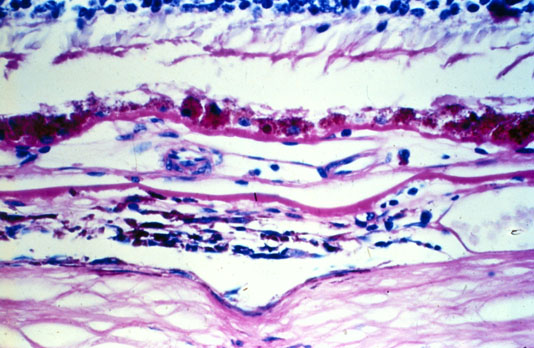
C. One of 6 sources of neovascularization from the choroid.

Figures 12C reprinted with permission, from: Chang TS, Freund KB, de la Cruz Z, Yannuzzi LA, Green WR. Clinicopathologic correlation of choroidal neovascularization demonstrated by indocyanine green angiography in a patient with retention of good vision for almost four years. Retina 1994; 14:114-24.