![]() Figure 11 of
Green, Mol Vis 1999;
5:27.
Figure 11 of
Green, Mol Vis 1999;
5:27.
Figure 11. Occult choroidal neovascularization
A. Red-free photograph of right macula showing subretinal fluid surrounded by lipid (arrows), overlying apparent choroidal neovascularization.
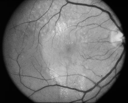
Figures 11A reprinted with permission, from: Bressler SB, Silva JC, Bressler NM, Alexander J, Green WR. Clinicopathologic correlation of occult choroidal neovascularization in age-related macular degeneration. Arch Ophthalmol 1992; 110:827-32.
B. Angiogram demonstrates occult choroidal neovascularization with staining of irregularly elevated area of retinal pigment epithelium and poorly demarcated leakage in the late phase in which the exact boundaries of the neovascularization cannot be determined with certainly.

Figures 11B reprinted with permission, from: Bressler SB, Silva JC, Bressler NM, Alexander J, Green WR. Clinicopathologic correlation of occult choroidal neovascularization in age-related macular degeneration. Arch Ophthalmol 1992; 110:827-32.
C. Area of fluorescein staining corresponds to a 100 µm-thick scar with a 10 µm non-vascularized subretinal and 90 µm vascularized intraBruch's membrane components. Periodic acid Schiff, x25.

Figures 11C reprinted with permission, from: Bressler SB, Silva JC, Bressler NM, Alexander J, Green WR. Clinicopathologic correlation of occult choroidal neovascularization in age-related macular degeneration. Arch Ophthalmol 1992; 110:827-32.
D. Area shows a single source of neovascularization (asterisk) from the choroid through a defect in Bruch's membrane (between arrows). Periodic acid Schiff, 40.

Figures 11D reprinted with permission, from: Bressler SB, Silva JC, Bressler NM, Alexander J, Green WR. Clinicopathologic correlation of occult choroidal neovascularization in age-related macular degeneration. Arch Ophthalmol 1992; 110:827-32.