![]() Figure 10 of
Green, Mol Vis 1999;
5:27.
Figure 10 of
Green, Mol Vis 1999;
5:27.
Figure 10. Occult choroidal neovascularization
A. Ophthalmoscopic appearance of area of pigment modeling contiguous with an inferior area of RPE detachment. B. Fluorescein angiographic appearance of lacy late staining superior and a serous RPE detachment inferiorly.

Figures 10A-B reprinted with permission, from: Small ML, Green WR, Alpar JJ, Drewry RE. Senile macular degeneration. A clinicopathologic correlation of two cases with neovascularization beneath the retinal pigment epithelium. Arch Ophthalmol 1976; 94:601-7.
C. Area where a choroidal vessels (asterisks) extends through a defect in Bruch's membrane and into a plane located between BlamD with RPE and the remainder of Bruch's membrane (Verfoeff van Geisen, x40)
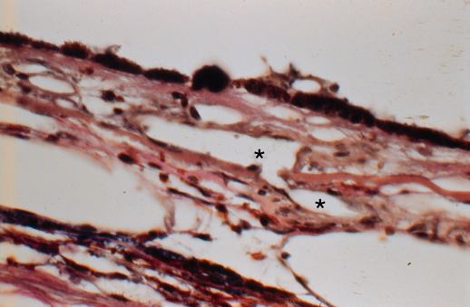
Figures 10C reprinted with permission, from: Small ML, Green WR, Alpar JJ, Drewry RE. Senile macular degeneration. A clinicopathologic correlation of two cases with neovascularization beneath the retinal pigment epithelium. Arch Ophthalmol 1976; 94:601-7.