![]() Figure 1 of
Green, Mol Vis 1999;
5:27.
Figure 1 of
Green, Mol Vis 1999;
5:27.
Figure 1. Flow diagram illustrates the relationships of the various morphologic forms of age-related macular degeneration
Age-related macular degeneration is a diffuse process that involves the RPE, photoreceptor cells and perhaps the choroidal vasculature. The first morphologic features is the development of basal deposits. Unfortunately, this phase only becomes evident with the effects on adjacent tissues including: pigment mottling with RPE clumping, attenuation, hypertrophy, hyperplasia, and atrophy; soft drusen; RPE detachment; and choroidal neovascularization. Choroidal neovascularization may then lead to serous and/or hemorrhagic detachment of the RPE and/or the neurosensory retina, pigment mottling, exudation, hemorrhage, and RPE tears. These features lead to a reparative process--a disciform scar that usually has subretinal and sub-RPE components. Disciform scars may have vascular contributions from the retina, marked exudation, and contribute to RPE tears.
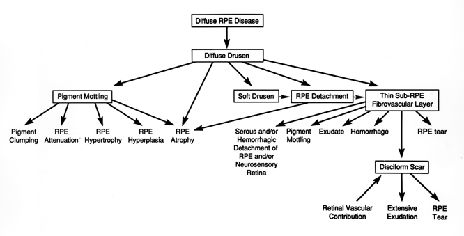
Figure 1 reprinted with permission, from: Green WR. Age-related macular degeneration. In: Franklin RM, editor. Retina and vitreous: proceedings of the Symposium on Retina and Vitreous, New Orleans, LA, USA, March 12-15, 1992. New York: Kugler; 1993. p. 7-13.