![]() Figure 4 of
de Peyer, Mol Vis 1999;
5:23.
Figure 4 of
de Peyer, Mol Vis 1999;
5:23.
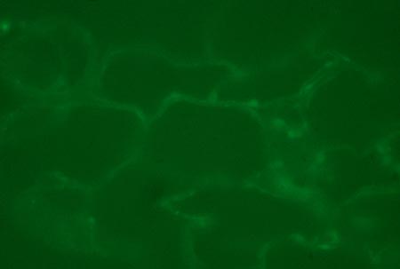
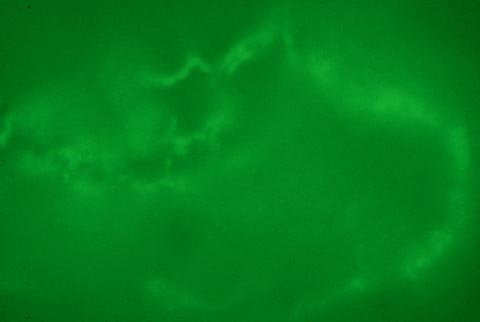
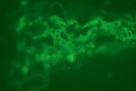
Figure 4. Immunohistochemical analysis of young leaf and root tissues
Some slides showed irregular cell structure since adhesion of the microtomed samples to the slides was poor; they had a tendency to lift off partially and present a distorted or corrugated appearance. This is an inherent drawback of plant tissues and we are aware of no superior plant immunohistochemical protocol for light microscopy. The maginification was x200 for all figures. The longest dimension of each image is approximately 180 µm. All our controls, including those with primary and secondary antibodies alone, were negative and showed no fluorescence.
A. Young transgenic leaf section showing enhanced immunofluorescence at cell peripheries. Young leaves were dissected, and fixed in 4% paraformaldehyde. Slides were dried, and primary anti-MIP antibody was added. Secondary antibody (anti-Rabbit FITC conjugate) was then added. Propidium iodide nuclear stain (3 µg/ml) was then briefly applied, coverslips were mounted on the slides and sealed with nail varnish.
B. Young transgenic leaf section showing enhanced immunofluorescence at cell peripheries. Young leaves were dissected, and fixed in 4% paraformaldehyde. Slides were dried, and primary anti-MIP antibody was added. Secondary antibody (anti-Rabbit FITC conjugate) was then added. Propidium iodide nuclear stain (3 µg/ml) was then briefly applied, coverslips were mounted on the slides and sealed with nail varnish.
C. Young leaf section from control plant showing background immunofluorescence. Young leaves were dissected, and fixed in 4% paraformaldehyde. Slides were dried, and primary anti-MIP antibody was added. Secondary antibody (anti-Rabbit FITC conjugate) was then added. Propidium iodide nuclear stain (3 µg/ml) was then briefly applied, coverslips were mounted on the slides and sealed with nail varnish. From our numerous plant (and indeed root) cell sections, we have always seen some background immunofluorescence localized to cell membranes.