![]() Figure 5 of
Hawes, Mol Vis 1999;
5:22.
Figure 5 of
Hawes, Mol Vis 1999;
5:22.
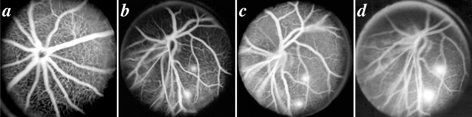
Figure 5. Fluorescein angiograms
A: Normal C57BL/6J mouse eight weeks of age, taken in early venous phase. All retinal arterioles and venules are filled with dye and details of the retinal capillary bed are easily visualized. B-D: Eight week old Onc1 mouse, demonstrating an abnormal blood vessel pattern and two areas of inferolateral fluorescein leakage that become more apparent from early to late venous stages. The angiograms in B-D were taken 143, 183, and 209 s after fluorescein administration.
For a larger version of an individual image, click on the appropriately named text link below.
A - B - C - D