![]() Figure 4 of
Hawes, Mol Vis 1999;
5:22.
Figure 4 of
Hawes, Mol Vis 1999;
5:22.
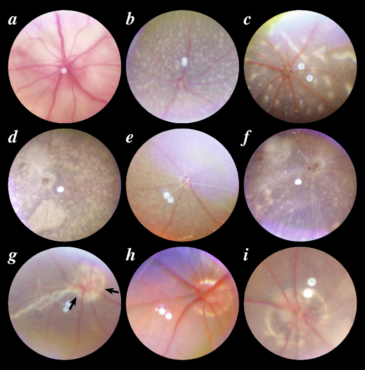
Figure 4. Catalogue of mutant phenotypes caused by the mouse mutations listed
Ages were typically selected to demonstrate well developed retinal changes. A: Achondroplasia (cn) at 6 weeks shows dilated venules. B: Typical retinal degeneration 7 (rd7) 3 weeks of age demonstrates a pattern of small white spots. C: In atypical rd7 at 4 weeks of age, spots are larger, often elongated and fewer in number. D: Nervous (nr) 27 weeks of age shows larger spots that are forming confluent plaques of RPE hypopigmentation. All retinal vessels show severe narrowing. E: Motor neuron degeneration (mnd), 12 weeks of age, demonstrates retinal arteriole narrowing on a background of faintly hypopigmented spots. F: Purkinje cell degeneration (pcd), 45 weeks of age, also has severe narrowing of all retinal vessels. Hypopigmented spots of variable size and irregular arrangement are evident. G: Kidney and retinal defects (Krd), 12 weeks of age, shows a large coloboma of the optic nerve (arrows) and a sectoral chorioretinal coloboma. H: Mice with the optic nerve coloboma phenotype (ONC), four weeks of age demonstrates a large coloboma of the optic nerve. I: CALB/Rk, 19 weeks of age, has a large optic nerve coloboma that appears to include peripapillary retina.
For a larger version of an individual image, click on the appropriately named text link below.
A - B - C