![]() Figure 2 of
Zimmerman, Mol Vis 1999;
5:19.
Figure 2 of
Zimmerman, Mol Vis 1999;
5:19.
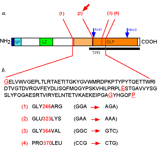
Figure 2. Structure of TIGR protein and mutations evaluated in the translocational assay
A. Important sequence motifs are shown: SP=signal peptide, LZ=leucine zipper, OLF=olfactomedin homology domain. Two alternative start sites for the SP were predicted; the region using the first start site is indicated by dark blue, and the second by light blue. The region of olfactomedin homology used by Rozsa et al [5] is shown in orange. The region of higher homology shown in dark orange, and lower homology in light orange. The translocational paused region=TPR, determined according to Methods, and is indicated by the black bar. Restriction enzymes sites in the translocational pausing assay used to generate the truncated forms of TIGR are indicated in blue with arrows. The mutations tested are numbered 1-4, with the E323K mutation marked with a red arrow. B. The TIGR protein sequence is shown, with the location of mutations evaluated in this study highlighted in red.