![]() Figure 7 of
Park, Mol Vis 1999;
5:18.
Figure 7 of
Park, Mol Vis 1999;
5:18.
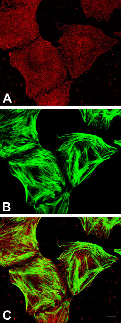
Figure 7. Immunolocalization of PLC-[gamma]1 and F-actin
CEC maintained in DMEM-10 were treated with cytoskeleton stabilization buffer prior to being subjected to immunofluorescent analysis, fixed, permeabilized and stained in the solutions containing the cytoskeleton stabilization buffer. F-actin staining was achieved with fluorescein-conjugated phalloidin. Texas Red signals are PLC-[gamma]1-positive (A); fluorescein signals are F-actin-positive (B); these two signals were digitally overlaid in C. No colocalization of PLC-[gamma]1 and F-actin is observed. Data are representative of six experiments. Bar, 5 µm.