![]() Figure 5 of
Park, Mol Vis 1999;
5:18.
Figure 5 of
Park, Mol Vis 1999;
5:18.
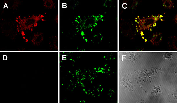
Figure 5. Colocalization of GST-SH3 and vinculin
CEC were microinjected with GST-SH3. Two h after injection, cells were treated with FGF-2 (10 µg/ml) supplemented with heparin (10 µg/ml) for an additional 4 h. Cells were then fixed, permeabilized, and stained with anti-PLC-[gamma]1 antibody and anti-vinculin antibody as described in the text. Red signals are PLC-[gamma]1-positive (A); green signals are vinculin-positive (B); yellow signals show the colocalization of SH3 domain and PLC-[gamma]1 (C). The same cell was further analyzed using confocal optical sectioning (D and E); GST-SH3 signal was present in the superficial layer (A) but was lost toward the basal membrane (D), while vinculin signal was maintained throughout the cell depth (B and E). F, phase-contrast image. Data are representative of three microinjection experiments. Bar, 10 µm.