![]() Figure 6 of
Ko, Mol Vis 1999;
5:17.
Figure 6 of
Ko, Mol Vis 1999;
5:17.
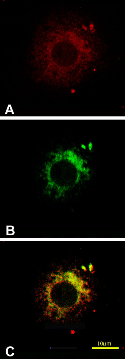
Figure 6. Immunofluorescent localization of procollagen I and prolyl 4-hydroxylase in CEC
Cells were fixed, permeabilized, and stained as described in the text. A. CEC stained for procollagen I. B. CEC stained for prolyl 4-hydroxylase; C. Superimposed signals of A and B. Bar, 10 µm.