![]() Figure 3 of
Ko, Mol Vis 1999;
5:17.
Figure 3 of
Ko, Mol Vis 1999;
5:17.
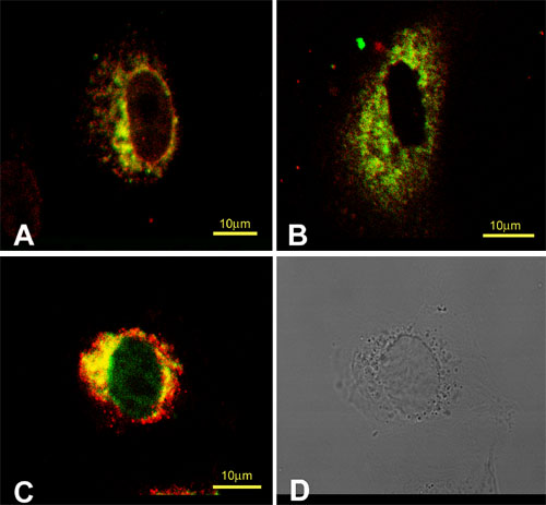
Figure 3. Immunofluorescent localization of procollagen I and Hsp47 in the presence or absence of brefeldin A treatment in CEC
Cells were treated with 2 µg/ml brefeldin A for 30 min, fixed, permeabilized, and stained as described in the text. A. Procollagen I (green) and Hsp47 (red) in brefeldin A-treated cells. B. Prolyl 4-hydroxylase (green) and procollagen I (red) in brefeldin A-treated cells. C. Prolyl 4-hydroxylase (green) and Hsp47 (red) in brefeldin A-treated cells. D. Phase-contrast microscopy of C. Bar, 10 µm.