![]() Figure 2 of
Ko, Mol Vis 1999;
5:17.
Figure 2 of
Ko, Mol Vis 1999;
5:17.
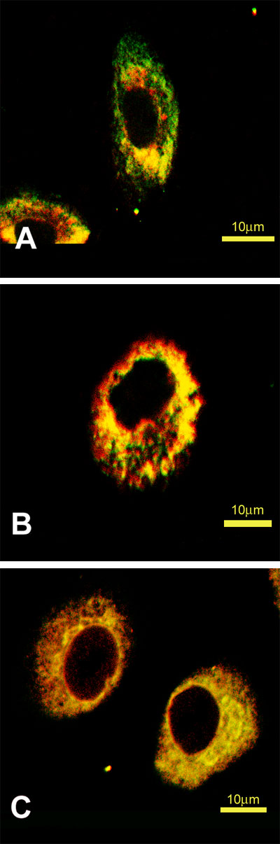
Figure 2. Immunofluorescent localization of procollagen I and Hsp47 in the presence or absence of [alpha],[alpha]'-dipyridyl treatment in CEC
Cells were treated with 0.3 mM [alpha],[alpha]'-dipyridyl for 2 h. Cells were then fixed, permeabilized, and stained as described in the text. A. Control cells stained with fluorescein (green) for procollagen I and with Texas-red (red) for Hsp47. B. Procollagen I (green) and Hsp47 (red) in [alpha],[alpha]'-dipyridyl-treated cells. C. Prolyl 4-hydroxylase (green) and procollagen I (red) in [alpha],[alpha]'-dipyridyl-treated cells. Bar, 10 µm.