![]() Figure 1 of
Ko, Mol Vis 1999;
5:17.
Figure 1 of
Ko, Mol Vis 1999;
5:17.
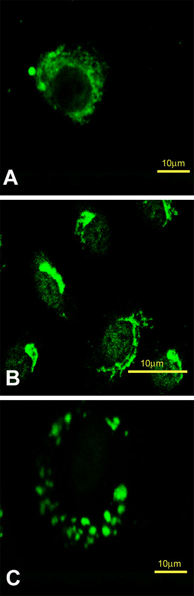
Figure 1. Distribution of markers for the ER, Golgi and lysosomes in CEC
Cells were prepared as described in the text, fixed, permeabilized, and stained with anti-chick prolyl 4-hydroxylase antibody for the ER, anti-Golgi 58K protein antibody for Golgi, and anti-LAMP-2 antibody for lysosome. A. ER staining. B. Golgi staining. C. Lysosome staining. Bar, A and B, 5 µm; C, 10 µm.