![]() Figure 4 of
Ayyagari, Mol Vis 1999;
5:13.
Figure 4 of
Ayyagari, Mol Vis 1999;
5:13.
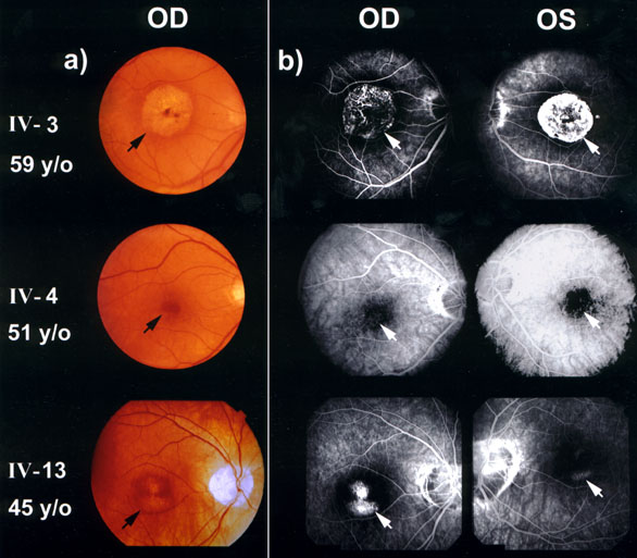
Figure 4. Clinical examination
Fundus photographs A and fluorescein angiograms B on affected males
Individual IV-3: Both maculae have extensive RPE atrophy and choriocapillary loss across the central three disc diameters that is readily apparant in the color photograph and the angiograms (early phase right eye and late phase left eye).
Individual IV-4: Both maculae show circumscribed thinning of the RPE across the central one disc diameter that gives a reddish hue in the color photograph and a subtle RPE "window defect" on the angiograms.
Individual IV-13: Bilateral macular atrophy is evident.