![]() Figure 2 of
Kim, Mol Vis 1999;
5:12.
Figure 2 of
Kim, Mol Vis 1999;
5:12.
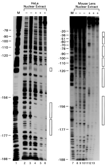
Figure 2. DNase I footprinting analysis of mouse MIP gene sequence -213/+71, with mouse and HeLa nuclear extract.
A [32P] 5'-end-labeled NcoI/EcoN1 DNA fragment corresponding to mouse MIP sequence -215/+71 was incubated with 15 µg lens (lanes 8-13) or HeLa (lanes 2-6) nuclear extracts. The reaction mixtures were then digested with DNase I: 0.3 units [lanes 4,11], 1 unit [lanes 5,12], and 3 units [lanes 6 and 13]. Protected regions are indicated with empty boxes to the right of each autoradiogram. M (lanes 1 and 7), G+A Maxam-Gilbert sequencing ladder. Mouse MIP gene 5'-flanking sequence present in the corresponding DNA fragments are indicated to the left.