![]() Figure 3 of
Farboud, Mol Vis 1999;
5:11.
Figure 3 of
Farboud, Mol Vis 1999;
5:11.
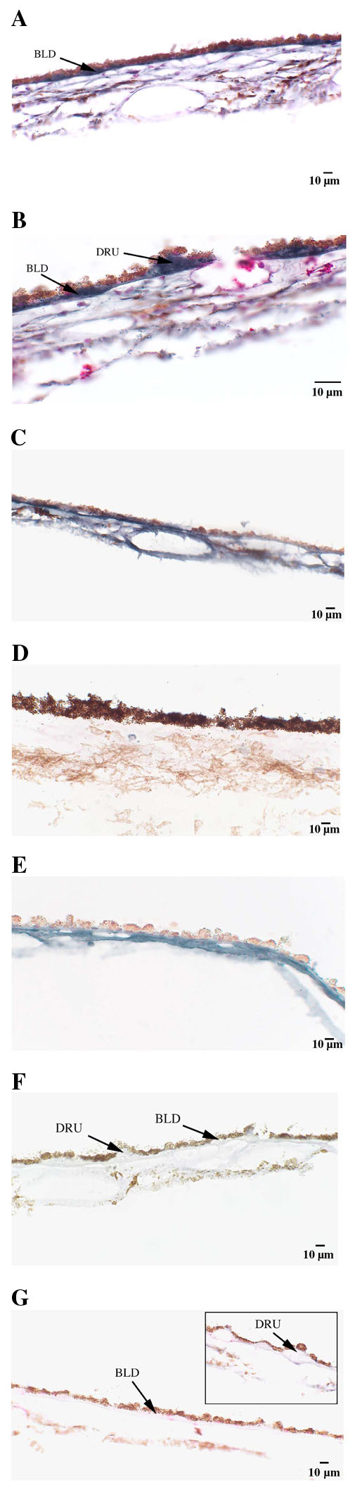
Figure 3. Immunohistochemical location of AGEs in human RPE-Bruch's membrane-choroid.
A. An 82 year old nondiabetic eye with staining in Bruch's membrane, basal laminar deposits, and choroid. B. Drusen also stained with the anti-AGE antibody. C. An 82 year old diabetic eye showed staining of AGEs in Bruch's membrane and choroid. D. In contrast, a 20 month old nondiabetic donor showed no staining. E. Significant staining is seen in the 82 year old nondiabetic eye after preincubating anti-AGE antibody with excess CML-BSA and pentosidine-BSA. F. The anti-AGE antibody preincubated with excess AGE-BSA in the 82 year old nondiabetic eye shows minimal staining. G. Minimal reaction was observed with nonimmune rabbit IgG. Key: Basal laminar deposit (BLD) and drusen (DRU).