![]() Figure 1 of
Koenekoop, Mol Vis 1999;
5:10.
Figure 1 of
Koenekoop, Mol Vis 1999;
5:10.
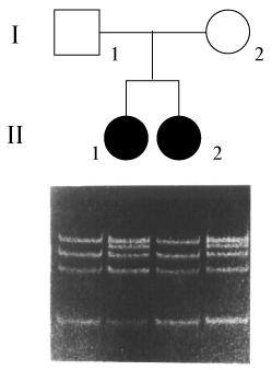
Figure 1. Example of a restriction diagnostic digest for the Tyr321Tyr polymorphism
The more frequent human allele has MaeIII sites that generate bands of 136, 115 and 96 bp (a 14 bp fragment cannot be seen) as shown in the first and third lanes from left to right. A homozygote for the Tyr321Tyr polymorphism (with one MaeIII site destroyed) would have bands of 136, 129 and 96 bp. A heterozygote has bands at 136, 129, 115, and 96 bp (as shown in the second and fourth lanes on the gel image). An example from a Greek LCA pedigree shows that one affected sib is heterozygous for the polymorphism (second lane), the other has two copies of the common allele (third lane). Lanes in the image correspond to each of the family members of the pedigree (lane one, the father; lane two, affected sib number 1; lane three, affected sib number 2, and lane four, the mother). The low molecular weight band in each lane is compatible with primer dimer formation.