![]() Figure 4 of
Sergeev, Mol Vis 4:9, 1998.
Figure 4 of
Sergeev, Mol Vis 4:9, 1998.
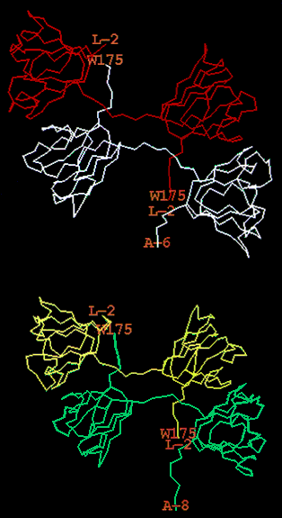
Figure 4. Terminal extensions of the bovine ßB2-crystallin structure
The location of N- and C-terminal arms in two ßB2-crystallin dimers from the asymmetric unit of the crystal were determined by X-ray crystallography [4]. The asymmetric unit contains two dimers appeared to form a tetramer. Molecule A of the first dimer is white, the molecule B is red and molecules C and D from the second dimer are yellow and green, respectively. Positions of Leu-2 and Trp175 are labelled as L-2 and W175, respectively.