![]() Figure 5 of
Singh, Mol Vis 4:7, 1998.
Figure 5 of
Singh, Mol Vis 4:7, 1998.
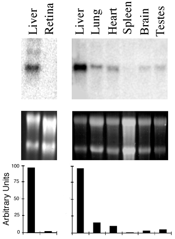
Figure 5. Multitissue Northern blot analysis of mouse PEDF mRNA.
The northern blot was performed as described in Methods. The upper panel shows the autoradiogram of the probed blot, the middle panel is the SYB green II stained gel, and the lower panel shows the relative PEDF mRNA levels normalized to the ribosomal 18S band.