![]() Figure 1 of
Hopp, Mol Vis 4:5, 1998.
Figure 1 of
Hopp, Mol Vis 4:5, 1998.
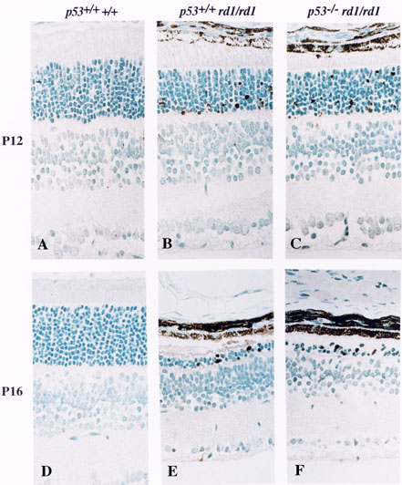
Figure 1. Representative central retinal cross sections from p53+/+ +/+, p53+/+ rd1/rd1, and p53-/- rd1/rd1 mice at P12 and P16.
Panels A-F compare the retinal morphology observed at P12 and P16 in normal mice and those of the p53+/+ rd1/rd1 and p53-/- rd1/rd1 genotypes. Panels B and C illustrate the slight increases in TUNEL label in rd1 mutant mouse retinas, regardless of p53 status, as compared to normal mice (A) at this early stage of the degeneration. Panels E and F demonstrate the loss of photoreceptor nuclei in the outer nuclear layer (ONL) induced by the rd1 retinal degeneration only 4 days later in the presence or in the absence of the p53 gene, respectively. Panel D shows a normal retina at P16. The absence of the p53 gene does not protect the rd1 mutant retina from apoptosis. Retinas from p53-/- +/+ and p53+/+ +/+ are indistinguishable at all ages (not shown). Paraffin sections were labeled by the TUNEL assay [11] and counterstained with methyl green (total magnification: x380).