![]() Figure 6 of
Nickerson, Mol Vis 4:33, 1998.
Figure 6 of
Nickerson, Mol Vis 4:33, 1998.
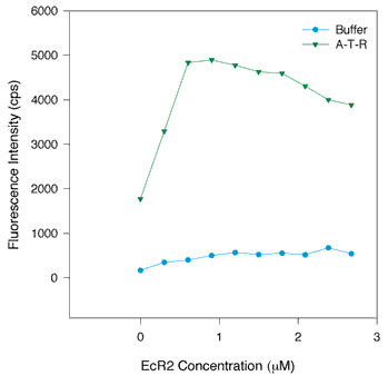
Figure 6. The binding of EcR2 to retinol: Titration of the protein-retinol interaction by varying the protein concentration
The Kd measured by increasing the retinol concentration at a constant protein concentration should be the same as the Kd found when varying the receptor (protein) concentration while holding the retinol concentration constant. The green triangles represent 1 µM solution of all-trans-retinol to which EcR2 was added. The blue circles represent the addition of EcR2 to buffer containing no retinol. Inspection of the plot suggests that the Kd measured by adding small increments of protein is the same as the Kd measured by incrementing the retinol concentration.