![]() Figure 9 of
Baer, Mol Vis 4:30, 1998.
Figure 9 of
Baer, Mol Vis 4:30, 1998.
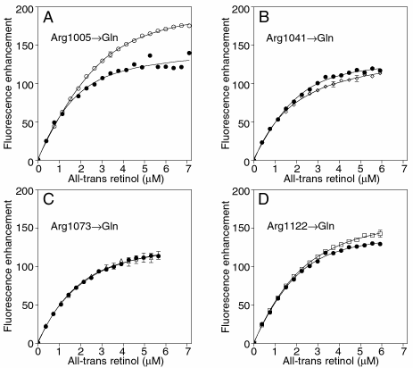
Figure 9. Binding of all-trans retinol to X4IRBP and the Arg->Gln mutants followed by monitoring ligand fluorescence enhancement upon binding (excitation 325 nm; emission 480 nm)
Each panel is a separate experiment comparing a single Arg->Gln mutant (open symbols) to that of X4IRBP (filled symbols). The binding curves are similar except for the Arg1005->Gln mutant where more ligand was required to reach saturation and the level of fluorescence enhancement attained was higher (panel A). Protein concentration was 1 µM.