![]() Figure 8 of
Baer, Mol Vis 4:30, 1998.
Figure 8 of
Baer, Mol Vis 4:30, 1998.
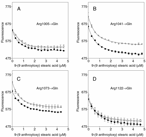
Figure 8. Binding of 9-AS to X4IRBP and the four Arg->Gln mutants monitoring quenching of protein fluorescence (excitation 280 nm; emission 340 nm)
Each panel is a separate experiment comparing a single Arg->Gln substitution (open symbols) to that of X4IRBP (filled symbols). Note that the shape of the quenching curves is similar for X4IRBP and the mutants. Protein concentration was 1 µM.